Plant Cell Under Light Microscope Labeled : Cells Cells To Systems Ks3 Biology Revision Bbc Bitesize - Plant cell under the microscope.. Make sure your straight labelling lines match the label exactly! Write down the magnification power of the objective lens. The structure that can be observed under the light microscope is. Image:plant cell seen under electron microscope. You know what, the onion cells look like bricks of a parapet wall when you see it under the low power of microscope.
Under a light microscope, the cell membrane, nucleus and cytoplasm of a cheek cell (animal cell) can be observed. In truth, there are still features of. Plant cell features (light microscope). The structure that can be observed under the light microscope is. It also has a very high resolving power.
What Is A Diagram Of A Plant And Animal Cell Under An Electron Microscope Quora from qph.fs.quoracdn.net A compound light microscope is a microscope with more than one lens and its own light source. A cell is a very tiny structure which exists in living bodies. A scanning electron microscope (sem) is a type of electron microscope that produces images of a sample by scanning the surface with a focused beam of electrons. Plant cell under the microscope. Students can print images to help them learn the cell. Winter jasmine leaf under a microscope (leaf of winter jasmine c. But at the same time it is interpretive. Under a light microscope, the cell membrane, nucleus and cytoplasm of a cheek cell (animal cell) can be observed.
A cell is a very tiny structure which exists in living bodies.
But at the same time it is interpretive. Students will finish plant cell diagrams from monday. When we look at cells under the microscope, our usual measurements fail to work. Light microscopes using visible light and lenses to form a magnified image of the object under investigation e.g. I am isolating cell wall of tobacco cells. Winter jasmine leaf under a microscope (leaf of winter jasmine c. In most plant cells, the organelles that are visible under a compound {light} microscope are the cell wall, cell membrane, cytoplasm, central vacuole, and here's a diagram of a plant cell: Even though the overall length of a dna molecule is about 2 inches, it is not possible to see dna through light microscopy as the dna is present inside the nucleus inside the. This section on microscopy is meant as an introduction as learners will need. Plug in the microscope and turn on the light source. Each organelle needs to be clearly labelled and with each label you need to add a description of the function of that observing cells under a microscope. You could use immunohistochemical methods which combine the specificity of antibody probes to their antigens (i.e. Make a drawing of one onion cell, labeling.
Make sure your straight labelling lines match the label exactly! It also has a very high resolving power. When we look at cells under the microscope, our usual measurements fail to work. Plant cells have a fixed shape. Tulip stem cells at the.
Plant Cell Science Diagram Clipart Set 300 Dpi School Etsy In 2021 Plant Cell Diagram Science Diagrams Plant Cell from i.pinimg.com Light microscopes use a number of lenses to produce an image that can be viewed directly at the eyepiece. A few cell organelles can be seen when a plant cell is viewed under a light microscope. Light microscopes (also known as optical microscopes) are the for example, iodine is often used to stain plant cells because it colours the starch stored within the cells a blue the components are labelled with fluorescent tags, then laser light is shone at the sample. A cell is a very tiny structure which exists in living bodies. Labelled diagram of a plant cell under microscope posted on march 18 2011 by admin onion cells stained with methylene blue look at the images of onion cells as they would be seen under a microscope draw each magnification label appear high picture plant and animal cell … Plant cells contain many organelles such as ribosomes, the nucleus, the plasma membrane, the cell wall, mitochondria, and chloroplasts. You could use immunohistochemical methods which combine the specificity of antibody probes to their antigens (i.e. Once slides have been prepared, they can be examined under a microscope.
View plant cells under a microscope.
Write down the magnification power of the objective lens. Make a drawing of one onion cell, labeling. Plug in the microscope and turn on the light source. Plant cell features (light microscope). Plant cells usually have all the bits that animal cells have, plus a few extra things that animal cells don't have. Cells of plant or animal tissue. The structures within the cell are. A compound light microscope is a microscope with more than one lens and its own light source. Peroxisomes are involved in the process of photorespiration. Labelled diagram of a plant cell under microscope posted on march 18 2011 by admin onion cells stained with methylene blue look at the images of onion cells as they would be seen under a microscope draw each magnification label appear high picture plant and animal cell … When you look at animal or plant cells under the electron microscope, you. A scanning electron microscope (sem) is a type of electron microscope that produces images of a sample by scanning the surface with a focused beam of electrons. Photorespiration is the method by which plant cells respirate, taking up light and producing energy.
I am isolating cell wall of tobacco cells. Compound light microscope · explain why objects must be centered in the field of view before plant and animal cells lab objectives:. Make a drawing of one onion cell, labeling. Write down the magnification power of the objective lens. Resolving power is the ability to distinguish between separate things which are close to each other.
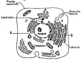
The Figure Below Is A Fine Structure Of A Generalized Animal Cell As Seen Under An Electron Microscope from www.kenyaplex.com Write down the magnification power of the objective lens. The structures within the cell are. A scanning electron microscope (sem) is a type of electron microscope that produces images of a sample by scanning the surface with a focused beam of electrons. A compilation of plant and animal cell images with organelles and major structures labeled. You could use immunohistochemical methods which combine the specificity of antibody probes to their antigens (i.e. Peroxisomes are involved in the process of photorespiration. Plant cells contain many organelles such as ribosomes, the nucleus, the plasma membrane, the cell wall, mitochondria, and chloroplasts. Plant cells have a fixed shape.
Light microscopes (also known as optical microscopes) are the for example, iodine is often used to stain plant cells because it colours the starch stored within the cells a blue the components are labelled with fluorescent tags, then laser light is shone at the sample.
It also has a very high resolving power. Under a light microscope, the cell membrane, nucleus and cytoplasm of a cheek cell (animal cell) can be observed. In this type of microscope, there are ocular lenses in the binocular eyepieces and objective lenses in a rotating nosepiece closer to the specimen. You know what, the onion cells look like bricks of a parapet wall when you see it under the low power of microscope. Plant and animal cells have a nucleus inside the cytoplasm. Polysaccharides) through the use of a typical fluorescence microscope? Observe the onion cell under both low and high power. Tulip stem cells at the. These include the cell wall, cell membrane, nucleus, chloroplasts. Plant cell under the microscope. See how a generalized structure of an animal cell and plant cell look with labeled diagrams. Write down the magnification power of the objective lens. This section on microscopy is meant as an introduction as learners will need.
Labelled diagram of a plant cell under microscope posted on march 18 2011 by admin onion cells stained with methylene blue look at the images of onion cells as they would be seen under a microscope draw each magnification label appear high picture plant and animal cell … plant cell under microscope labeled. But at the same time it is interpretive.
Share :
Post a Comment
for "Plant Cell Under Light Microscope Labeled : Cells Cells To Systems Ks3 Biology Revision Bbc Bitesize - Plant cell under the microscope."


Post a Comment for "Plant Cell Under Light Microscope Labeled : Cells Cells To Systems Ks3 Biology Revision Bbc Bitesize - Plant cell under the microscope."